


Previous blog entry:07 Wireless charging of dermatology device "Skimager"
Next blog entry: 09 Raspberry Pi and multispectral LED ring powered from the Qi Wireless Power kit - towards open source dermascope
During the Wireless Power Challange I have implemented wireless charging of skin dermatology device "Skimager". Here I will try to give some medical explanation what this device is doing.
Skin cancer melanoma is the fifth most common type of cancer. Dermatologists distinguish different risk groups by age, gender, skin color, sunburn history, hair color, number of nevi (birthmarks), part of the body, personal disease history as well as melanoma occurrence in family. Early detection is an efficient way to diagnose and manage skin cancer.
Skin structure
The epidermis is the top layer of the skin mostly made of flat cells called squamous cells. Below the squamous cells deeper in the epidermis are round cells called basal cells. Cells called melanocytes are scattered among the basal cells. They are in the deepest part of the epidermis. Melanocytes make the pigment (color) found in skin. When skin is exposed to UV radiation, melanocytes make more pigment, causing the skin to darken, or tan. Melanin is the primary determinant of skin color.
Melanocytic nevus (nevomelanocytic nevus, nevocellular nevus): benign proliferation of melanocytes, the skin cells that make the brown pigment melanin. Hence, most nevi are brown to black. They are very common; almost all adults have at least one, usually more. They may be congenital or acquired (usually at puberty).
Dermis: The dermis is the layer under the epidermis. The dermis contains many types of cells and structures, such as blood vessels, lymph vessels, and sweat and sebum glands. Sebum is an oily substance that helps keep your skin from drying out. Sweat and sebum reach the surface of your skin through tiny openings called pores.
Tumors
In general, cancers are caused by damage to DNA. Melanomas are usually caused by DNA damage resulting from exposure to ultraviolet (UV) light from the sun. Cancer is an abnormal growth of cell in a human body. Normal cells grow and divide to form new cells as the body needs them. When normal cells become old or get damaged, they usually die, and new cells take their place. But sometimes this process goes wrong and new cells form when the body doesn’t need them, and old or damaged cells do not die as they should. The buildup of extra cells often forms a mass of tissue called a growth or tumor. Growths on the skin can be benign (not cancer) or malignant (cancer). Benign growths are not as harmful as malignant growths.
Melanoma is most common skin cancer that can originate in any part of the body that contains melanocytes. Melanoma stages:
0: The melanoma involves only the top layer of skin. It is called melanoma in situ.
I: The tumor < 1 mm thick, the surface may appear broken down. Or the tumor is 1...2 mm thick, and the surface is not broken down.
II: The tumor is 1...2 mm thick, and the surface appears broken down. Or, the thickness of the tumor is more than 2 millimeters, and the surface may appear broken down.
III: The melanoma cells have spread to nearby lymph node or to tissues nearby.
IV: Cancer cells have spread to the lung or other organs, skin areas, or lymph nodes far away from the original growth.
These tumors have very indistinct margins. By allowing the surgeon to correctly identify the true extent of the tumor, repeat surgery often is decreased.
In 2013 I listened to a talk by leading Swedish cancer researcher Dr. Katharina Svanberg. She told that newest studies show that within lifetime human body successfully fights with ca 20 tiny cancer formations that the person does not even notice.
Dermatoscopy
Dermatoscopy - Wikipedia, the free encyclopedia
Dermatoscopy (also known as dermoscopy or epiluminescence microscopy) is the examination of skin lesions with a dermatoscope.
Dermoscopy cameras usually magnify the image 10 x. Small, inexpensive hand-held devices such as the DermLite or DermScope illuminate a skin lesion with polarized light, thus greatly reducing specular reflection. However, diagnosis of the lesions imaged is still left up to the physician, which is subjective and therefore can vary significantly.
Doctors use so called ABCDE-rule to distinguish benign from malignant spots on skin. A-Asymmetry, B-Border, C-Color, D-Diameter, E- Elevation.
http://www.sun-protection-and-products-guide.com/ABCDE-rule-for-skin-cancer.html

Basically, any mole or growth that is CHANGING needs to be checked by a physician.
Multispectral imaging
Accuracy of the diagnosis also decreases for non-expert physicians who do not specialize in melanoma detection. Furthermore, the use of white light does not allow for accurate depth and chromophore analysis. Multispectral imaging provides additional depth and chromophore information and computer aided analysis of dermoscopy images can be more objective.
- Ultraviolet wavelength causes skin fluorescence in longer wavelength. One can measure fluorescence parameter (malignant). Ultraviolet and blue wavelengths penetrate only ski surface layer.
- Infrared wavelength penetrates skin up to 1.5 mm deep. Nevus is visible in blue but in infrared it is practically not visible. This means that it is on the skin surface and not deeply in the tissue.
- All nevi show increased eritrema (blood perfusion redness) index meaning that all pathologies are having higher blood perfusion.
- Bruises show increased bilirubin index. Bilirubin has strong absorption in blue.
- Melanin shows increased absorption at blue that gradually decreases through green, red and infrared colors.
At the University of Latvia non-invasive melanoma diagnosis using multispectral imaging are done in collaboration with the hospital and optical methods were verified by histological methods. In order not to upset the tested person name “Melanoma index” is replaced by “C43 indicator”.
Below are absorption spectra of skin chromphores and LED emission spectra.

There are 4 LEDs for each wavelength to make illumination uniform. Light from LEDs is sent through a linear polariser sheet. In front of camera is another polariser sheet oriented at 90 degrees. This is a powerful method to eliminate direct reflection from the surface. Skin under investigation is illuminated for 1 s with each color and photo is taken.

Below is image obtained in white light, red, green, blue, infra-red and ultraviolet illumination.
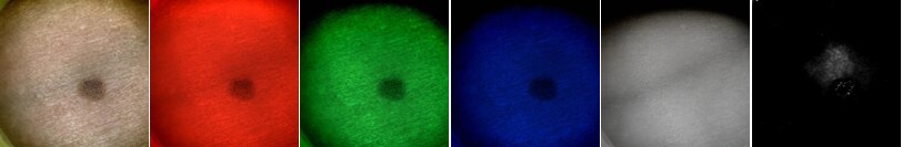
Such diagnostic information can be extracted and mapped on the original photo:
- Hemoglobin index R/G H=I660/I545 nm
- Bilirubin index B/R B=I450/I545
- Melanin index G/R/IR M=I545/I660/I940
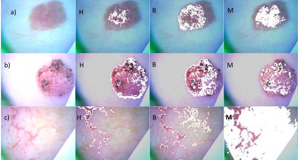
Picture below shows white light image and maps of Hemoglobin H, Bilirubin B, Melanin M for a) hemangioma, b) papilloma (wart), c) blood vessels.
Results are published in a recent article:
J. Spigulis, U. Rubins, E. Zaharans, J. Zaharans, L. Elste,
A device for multimodal imaging of skin
Proc. SPIE 8574, Multimodal Biomedical Imaging VIII, 85740J (March 13, 2013); doi:10.1117/12.2003510
Nice South Korea OptoBioMed promotional video about a similar working device:
https://www.youtube.com/watch?v=5Lpb6w2kotQ
Next blog entry: 09 Raspberry Pi and multispectral LED ring powered from the Qi Wireless Power kit - towards open source dermascope
Top Comments