


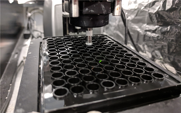
The bioprinting technology can print a 96-well array of human tissue samples in just 30 minutes. (Image Credit: Biofabrication)
Rapidly 3D printing large batches of customized biological tissues would ultimately make drug development faster and more affordable. Nanoengineers at the University of California San Diego developed a new bioprinting technology that 3D prints a 96-well array of human tissue samples in 30 minutes. The team says that rapidly producing samples like these could advance high-throughput preclinical drug screening and disease modeling.
A pharmaceutical company takes up to 15 years to produce a new drug, with costs reaching $2.4 billion. First, tens of thousands of drug candidates are screened in test tubes. Animals then get tested with any successful candidate, and the ones that pass this stage advance to clinical trials. If all goes well, a candidate becomes available to the market as an FDA-approved drug.
This process could be shortened with the newly developed high-throughput 3D printing technology. Drug developers could quickly produce large amounts of human tissues, which they could use to test and weed out drug candidates.
"With human tissues, you can get better data—real human data—on how a drug will work," said Shaochen Chen, a professor of nanoengineering at the UC San Diego Jacobs School of Engineering. "Our technology can create these tissues with high-throughput capability, high reproducibility and high precision. This could really help the pharmaceutical industry quickly identify and focus on the most promising drugs."
Even though animal testing could still exist, this technology would at least minimize failures during that process.
"What we are developing here are complex 3-D cell culture systems that will more closely mimic actual human tissues and that can hopefully improve the success rate of drug development," said Shangting You, a postdoctoral researcher in Chen's lab and co-first author of the study.
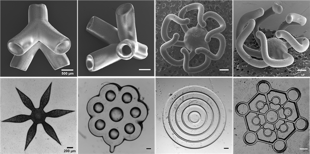
The bioprinting technology can rapidly print out these types of geometric shapes. (Image Credit: Biofabrication)
The technology has high-resolution printing capabilities. It produces lifelike human liver cancer tissues with blood vessel networks. Traditional techniques would take hours to print a tissue sample, but it only takes ten seconds with this method. One advantage is that it automatically prints samples in industrial well plates, eliminating the need to transfer them manually for screening.
The tissue samples produced with this technology are highly organized, allowing them to be replicated for industrial-scale screening. Chen explains that this is a different technique compared to growing organoids for drug screening. "With organoids, you're mixing different types of cells and letting them self-organize to form a 3-D structure that is not well controlled and can vary from one experiment to another. Thus, they are not reproducible for the same property, structure, and function. But with our 3-D bioprinting approach, we can specify exactly where to print different cell types, the amounts and the micro-architecture."
Using a computer, the team creates 3D models of biological structures to produce the tissue samples. Afterward, the computer splits the model into snapshots and sends them to millions of microscopic-sized mirrors. Every mirror projects violet light patterns in the snapshots. These light patterns are then beamed onto a solution with live cell cultures and light-sensitive polymers that turn solid when exposed to light. The sample is continuously printed one layer at a time, resulting in a 3D solid polymer scaffold encapsulating live cells that grow into biological tissue. The microscopic mirror array contributes to the printer's high speed due to 2D patterns being projected on the substrate while printing layer by layer.
The team automated this technology to perform high-throughput tissue printing. Allegre 3D licensed the technology and launched a commercial product.
Have a story tip? Message me at: http://twitter.com/Cabe_Atwell