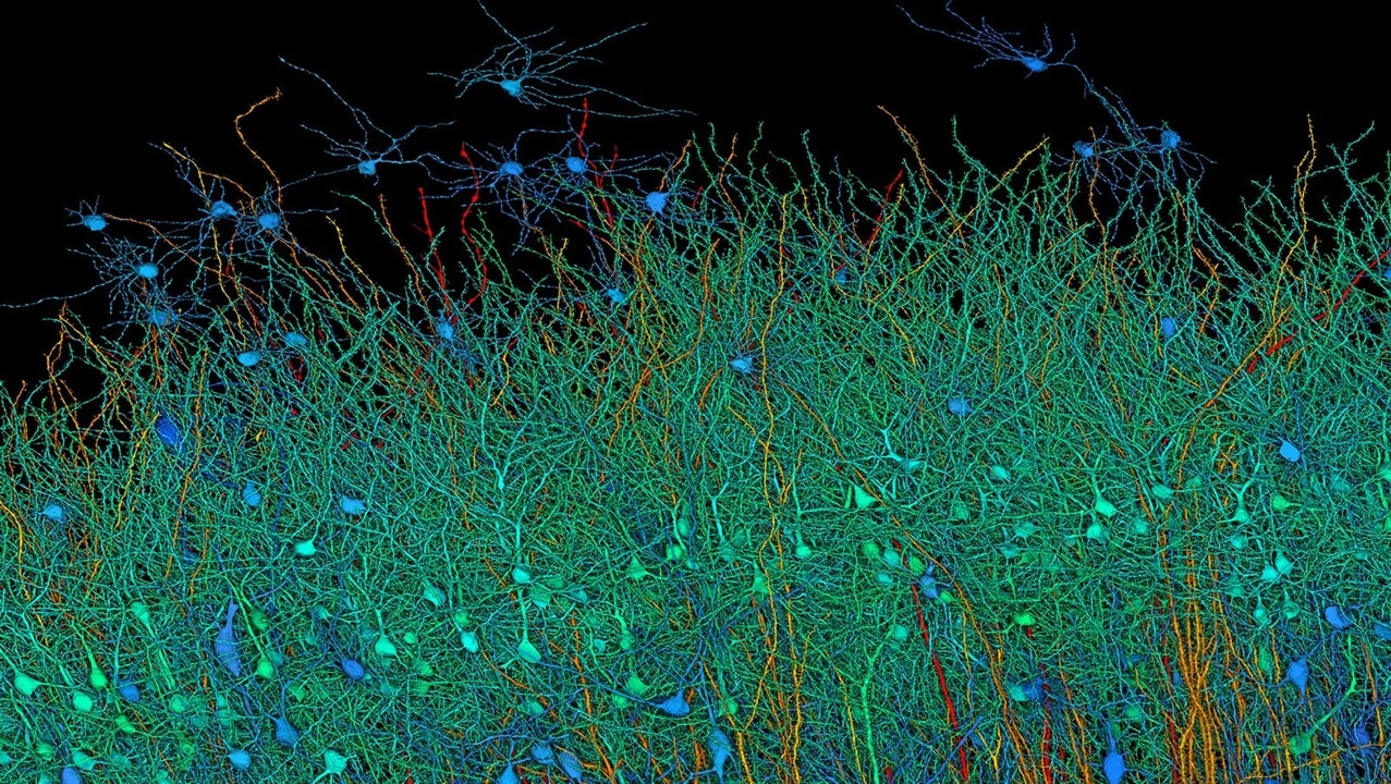
Researchers captured this image of a sample obtained from the anterior temporal lobe. (Image Credit: Google Research & Lichtman Lab (Harvard University). Renderings by D. Berger (Harvard University))
Researchers from Harvard and Google Research used an AI algorithm to produce a 3D map of a cubic millimeter of the human brain. The sample features 57,000 cells, 230 millimeters of blood vessels, and nearly 150 million synapses. To date, this is the highest-res image of the brain.
The researchers used Google’s machine learning tool to image the human brain, an approach that would’ve taken them a few years longer to complete without the tech. The team overcame a few obstacles before mapping it. For starters, they had to find a brain tissue sample, and cadaver tissue wouldn’t be ideal because the brain deteriorates rapidly after death. So, the researchers collected a tissue sample from a woman with epilepsy who underwent brain surgery to help maintain her seizures. After obtaining this sample, they preserved it in resin so they could slice it.
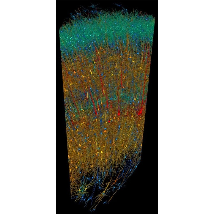
The researchers cut the brain sample into 5,000 slices that are thinner than human hair. From there, the team used a high-speed electron microscope to take photos of all the slices. However, the researchers faced another challenge: color-coding the wiring that traversed in all sorts of directions and had different connections. Google’s AI imaging model then came into play, precisely stitching those images back together, color-coding the wiring, and finding the connections. That process is difficult, and if they gave one wire the wrong color, each connection would be incorrect.

Wirings of the brain color-coded. (Image Credit: Google Research & Lichtman Lab (Harvard University). Renderings by D. Berger (Harvard University))
With these images, the team discovered some mind-boggling features. For example, one neuron had over 5,000 connection points with other neurons, cell clusters grew in replicated images of each other, and axons wound up in yarn ball-like shapes. The researchers were surprised by these findings, which aren’t in textbooks. Other researchers can access this map to discover more. Doing so allows them to study certain structures of the cortex in extreme detail to ensure everything is correct and publish their findings.
There are some impressive numbers worth looking into that can help us understand the brain sample size and data collected. A cubic millimeter of brain matter is one millionth the size of an adult human brain. Even then, the imaging and mapping of the sample represent 1.4 petabytes of data (layered TIFF files). Those scans would eat up 1.6 zettabytes if the AI completely mapped the human brain.
Have a story tip? Message me at: http://twitter.com/Cabe_Atwell
