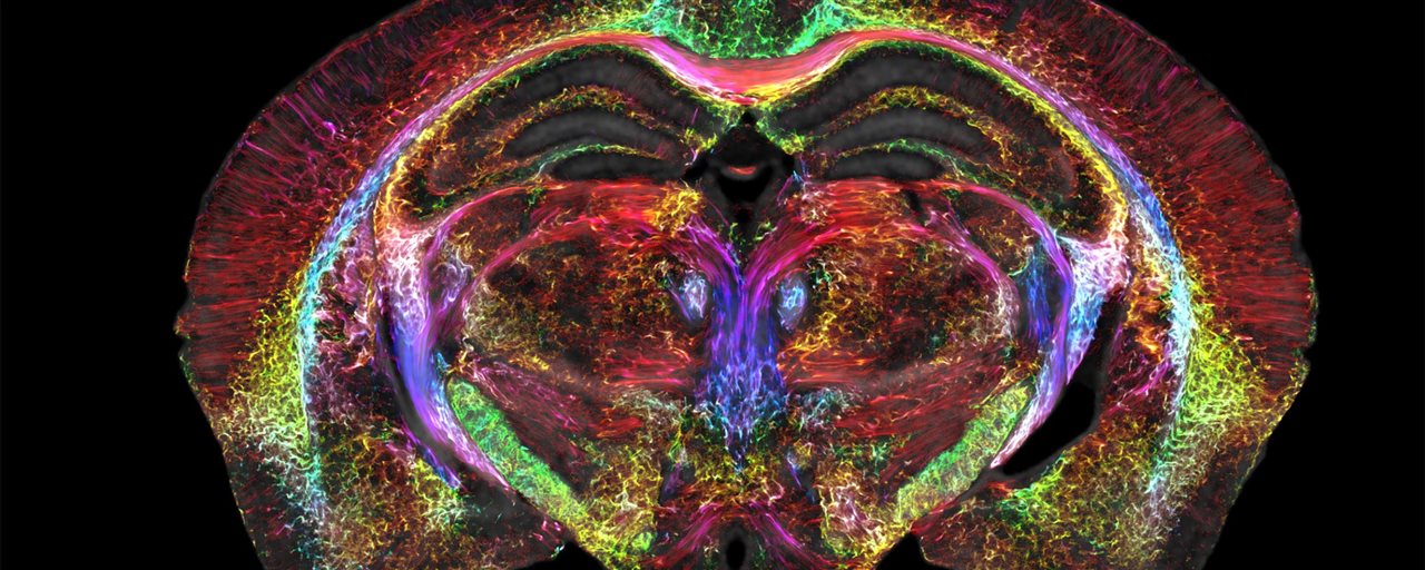
The researchers produced a colorful scan of a mouse's brain, allowing them to view neural network connections. (Image Credit: Duke Center for In Vivo Microscopy)
MRI imaging is useful for providing visualizations of soft tissue. However, MRI imagery isn't sharp enough to visualize microscopic details inside the brain even though it spots a tumor. Now after a decade-long effort, researchers at Duke University have improved MRI resolution to capture clearer brain images and potentially help scientists better understand neurodegenerative conditions.
Aligning with the 50th MRI anniversary, the team produced mouse brain scans that are significantly sharper compared to a traditional MRI. They described the improvement as "going from a pixilated 8-bit graphic to the hyper-realistic detail of a Chuck Close painting."
An individual voxel on the image measures five microns, which is 64 million times smaller than a clinical MRI voxel. While the researchers used the improved MRI on mice rather than humans, it still offers a way to visualize the brain's connections in crisp detail. According to the team, the data collected from mouse imaging can help scientists further understand how the brain changes with age, diet, or neurodegenerative diseases like Alzheimer's.
"It is something that is truly enabling. We can start looking at neurodegenerative diseases in an entirely different way," said G. Allan Johnson, Ph.D., the lead author of the new paper and the Charles E. Putman University Distinguished Professor of Radiology, physics and biomedical engineering at Duke.
While a clinical MRI uses a 1.5 to 3 Tesla magnet, Duke University's MRI has a 9.4 Tesla magnet, making it more powerful. It also features a set of gradient coils that assist with brain imaging and are 100 times stronger than clinical MRIs. Additionally, the refined MRI comes with one powerful computer that performs similarly to 800 fully running laptops to produce a brain image.
Once they complete a scan, the tissue undergoes light sheet microscopy imaging, a different technique that allows them to label specific groups of cells across the brain, including dopamine-issuing cells. Afterward, the team maps the light sheet images that provide very precise visualizations of brain cells. This step is achieved on the original MRI scan, which has a higher accuracy and shows a vivid visualization of circuits and cells throughout the brain.
All of that brain data imagery allows researchers to look into the brain and uncover mysteries in ways not attempted before. One set of images reveals how the brain connectivity changes as mice age and how the memory-involved subiculum changes more than the other regions of the mouse's brain. An additional set of images show rainbow-colored connections that represent the neural networks' deterioration in a mouse model with Alzheimer's.
With this highly-powered MRI, the team hopes it can lead to a better understanding of mouse models with human diseases like Huntington's and others. This would then enable them to uncover how these conditions function or worsen in patients.
"Research supported by the National Institute of Aging uncovered that modest dietary and drug interventions can lead to animals living 25% longer," Johnson said. "So, the question is, is their brain still intact during this extended lifespan? Could they still do crossword puzzles? Are they going to be able to do Sudoku even though they're living 25% longer? And we have the capacity now to look at it. And as we do so, we can translate that directly into the human condition."
Have a story tip? Message me at: http://twitter.com/Cabe_Atwell
